

liquid phase electron microscopy of bacterial macromolecular complexes in action
Project Leaders:
Dwayne Miller, Eike Schulz and Martin Aepfelbacher
Image proof:
Illustration: hegasy.de | © Infectophysics 2019
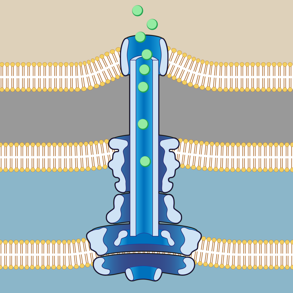
The type three secretion system (T3S) of pathogenic bacteria like Salmonella, Shigella and Yersinia is a multimolecular complex made up by 200 proteins and a total size of approximately 4 MDa. It is assembled in different phases and consists of several parts, i.e. a basal body embedded within the bacterial membranes and an extrabacterial needle like structure, which are distinguishable by conventional cryo electron microscopy. While static structures of T3S’s have been visualized by cryo EM in different states of assembly and in action, there are no recordings of the T3S dynamics beyond the resolution of light microscopy (around 200-400 nm). It’s a longtime dream to conduct electron microscopy (EM) as simple as light microscopy. Liquid phase EM (liquid EM) comes close to this dream.
In liquid EM the specimens are not dried or frozen, but retained in solution, which uniquely offers to observe not only the structure but also the motions of molecules in solution. These dynamic structure changes are key to a complete understanding of macromolecular function and therefore could also provide major new information as to the function of the bacterial type three secretion system.